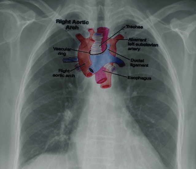
Aortic dissection is a condition of delamination of the aortic wall leading to the development of a double-barrel aorta compromised luminal and side branch flow and weakening of the aortic wall predisposing to aneurysmal degeneration. The first and largest branch that ascends laterally to split into the right common carotid and right subclavian arteries.
Right Aortic Arches Article
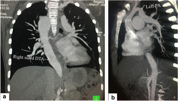
Right Sided Aortic Arch With Aortic Origin Of Left Pulmonary Artery And Patent Ductus Arteriosus A Rare Combination Of Aortic Arch Anomalies Springerlink
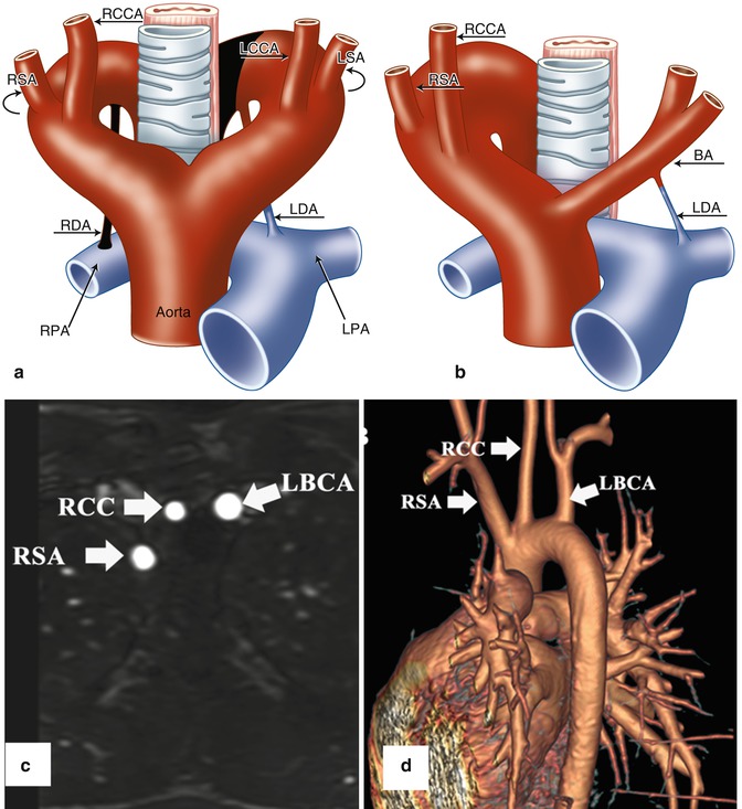
Aortic Arch Anomalies Radiology Key
Annals of Vascular Surgery.
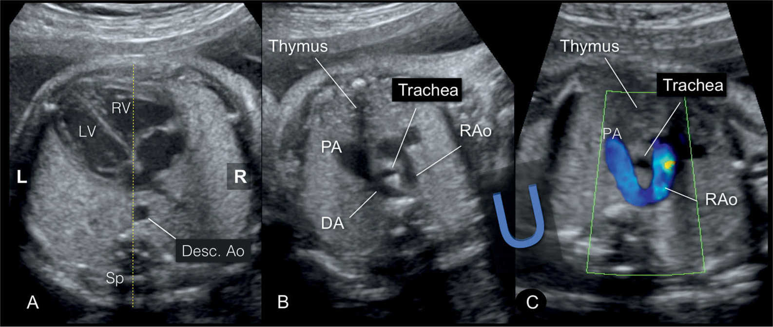
Right side aortic arch. Aberrant right subclavian arteries ARSA also known as arteria lusoria are among the commonest aortic arch anomalies. It often occurs when the weakened andor stiff left ventricle loses the ability to efficiently pump blood to the rest of the body. An oblique plane crossing the left ventricle C.
January 2016 - Recent advances in understanding and treating thoracic aortic disease November 2015 - Genetics of Cardiomyopathy September 2015 - Cardiovascular Disease in. The murmur can be heard best during systole since this is when blood is spraying out of the. Acute Kidney Injury and Renal Replacement Therapy After Fontan Operation.
The type of surgery will depend on the location and type of aneurysm and your overall health. The ascending aorta and underside of the aortic arch are replaced with a separate Dacron graft and the two grafts are connected together to. Thoracic aortic aneurysm open repair.
The sharpness of the angle can be different among individuals. Cusp orientation is identified by the adjacent left atrium LA and the right and left coronary arteries RCA LCA between commissures. Vascular rings are malformations of the aortic arch in the main blood vessel that leads from the heart.
TAVR is a revolutionary new heart valve treatment most commonly used to treat a tight aortic valve otherwise known as aortic stenosis TAVR stands for transcatheter aortic valve replacement it is also commonly referred to as TAVI which stands for transcatheter aortic valve implantation. TAVR and TAVI are the same thing. The journal serves the interest of both practicing clinicians and researchers.
Segmented left-sided heart components B. Learn more about APCs and our commitment to OA. Clinical presentation They are often asymptomatic but around 10 of peo.
A traversing view from the ascending aorta Asc Ao clearly visualized the aortic root cavity D. Figure 7 indicates a Stanford Type B aortic dissection arising distal to the left subclavian artery. A Sagittal oblique maximum-intensity-projection MR angiographic image shows aneurysm in mid ascending aorta arrow with normal-caliber aortic arch and descending thoracic aorta.
The International Journal of Cardiology is devoted to cardiology in the broadest senseBoth basic research and clinical papers can be submitted. New Journal Launched. In the blood supply of the heart the right coronary artery RCA is an artery originating above the right cusp of the aortic valve at the right aortic sinus in the heart.
An example of a vascular ring a double aortic arch is shown on the right. Most Cited Previous 3 Years Open Access. It usually requires emergency open chest surgery to repair or replace the first segment of the aorta where the tear started ascending aorta - the arch andor aortic valve.
This conditions is also know as cor pulmonale. The aortic arch gives rise to three arterial branches. The aortic arch is the part of the aorta between the ascending aorta and thoracic descending aorta.
There are three major branches arising from the aortic arch. This type of dissection occurs in the first part of the aorta closer to the heart and can be immediately life-threatening. Right aortic arch with mirror image branching is the second most common form of a right-sided arch after right arch with aberrant left subclavian artery.
A structure consisting of a curved top on two supports that holds the weight of something. Because of the malformation the aortic arch and its branches partly or completely encircle the windpipe trachea the esophagus or both. Stanford Type A Aortic Dissection.
The murmur of aortic stenosis is heard and felt loudest in the 2nd intercostal space on the right side Figure 5 this is the area directly over the aorta 13. The new surgical journal seeks high-quality case reports small case series novel techniques and innovations in all aspects of vascular disease including arterial and venous pathology trauma arteriovenous. Right-sided aortic arch is a type of aortic arch variant characterized by the aortic arch coursing to the right of the trachea.
Right-sided or right ventricular heart failure is defined as a process not a disease. Since the first issue was released in 1984 the goal of the journal has been to improve the management of patients with vascular diseases by publishing relevant papers that. Most commonly there is a larger dominant right arch behind and a smaller hypoplastic left aortic arch in front of the tracheaesophagus.
Right aortic arch with mirror image branching is strongly associated with CHD in up to 98 of cases including tetralogy of Fallot truncus arteriosus tricuspid atresia and transposition of the great arteries with. Rotational Thromboelastometry-Guided Transfusion Protocol to Reduce Allogeneic Blood Transfusion in Proximal Aortic Surgery With Deep Hypothermic Circulatory Arrest. Incidence Predictive Factors and Prognostic Impact of Right Ventricular Dysfunction Before Transcatheter Aortic Valve Implantation.
Anatomic Insights Regarding the Srivastavas Correction Factor for Calculating the Diameter of the Virtual Aortic Annulus from the Distance Between the Hinge Points of the Right and Non-coronary Cusps. For an ascending or aortic arch aneurysm a large cut incision may be made through the breastbone. These arteries supply the right side of.
The left and right main coronary arteries are subsequently reimplanted into the graft with fine permanent suture. Brief Reports and Innovations is a gold open access journal launched by Annals of Vascular Surgery. DAA is an anomaly of the aortic arch in which two aortic arches form a complete vascular ring that can compress the trachea andor esophagus.
Journal of Vascular Surgery is dedicated to the science and art of vascular surgery and aims to be the premier international journal of medical endovascular and surgical care of vascular diseases. It travels down the right coronary sulcus towards the crux of the heart. It supplies the right side of the heart and the interventricular septum.
International Journal of Cardiology is a transformative journal. Different configurations can be found based on the supra-aortic branching patterns with the two most common patterns being the right-sided aortic arch with mirror image branching and the right-sided aortic arch with aberrant left subclavian artery. Double aortic arch is a relatively rare congenital cardiovascular malformation.
Epidemiology The estimated incidence is 05-2. 5 36-year-old man with bicuspid aortic valve fused right and left cusps.
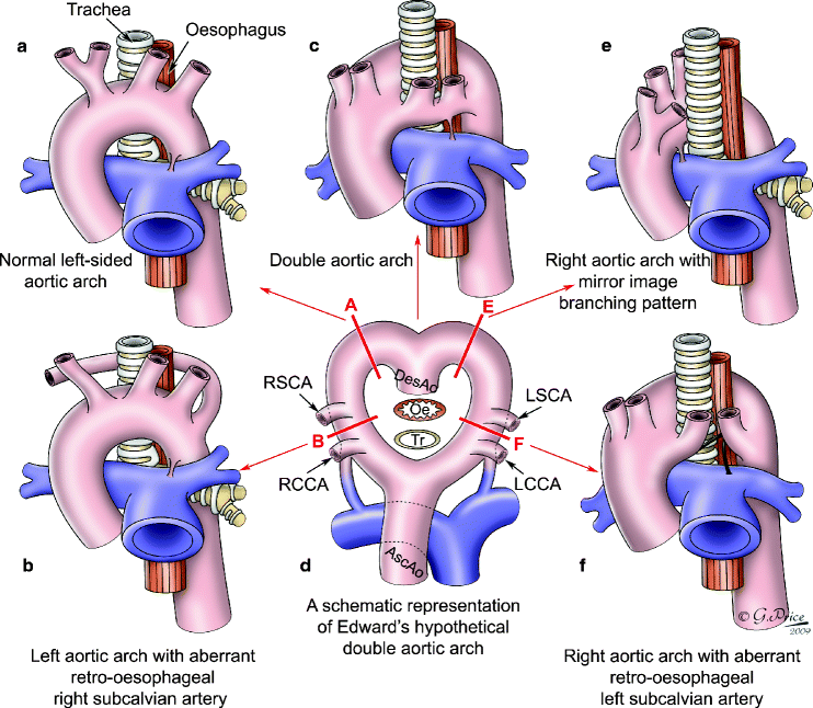
Aortic Vascular Rings Springerlink

Right Sided Aortic Arch Radiology Case Radiopaedia Org

Right Sided Aortic Arch Is A Type Of Aortic Arch Variant Grepmed

Imaging Findings In The Right Aortic Arch With Mirror Image Branching Of Arch Vessels An Unusual Cause Of Dysphagia Singh G Kharat A Sehrawat P Kulkarni V Med J Dy Patil

Right Aortic Arch Double Aortic Arch And Aberrant Subclavian Artery Obgyn Key

Right Aortic Arch With Aberrant Left Subclavian Artery Pediatric Radiology Reference Article Pediatricimaging Org Pedsimaging
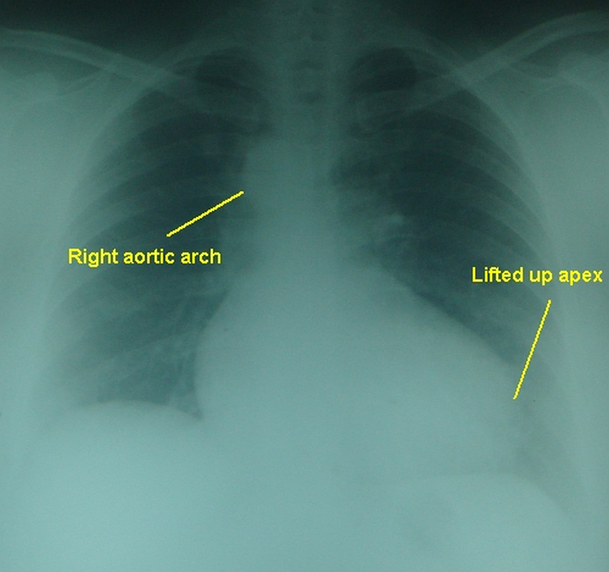
Tetralogy Of Fallot Right Aortic Arch All About Cardiovascular System And Disorders
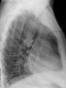
Right Sided Aortic Arch Wikipedia

